Celebrate Your Journey with a Maternity Ultrasound Scan
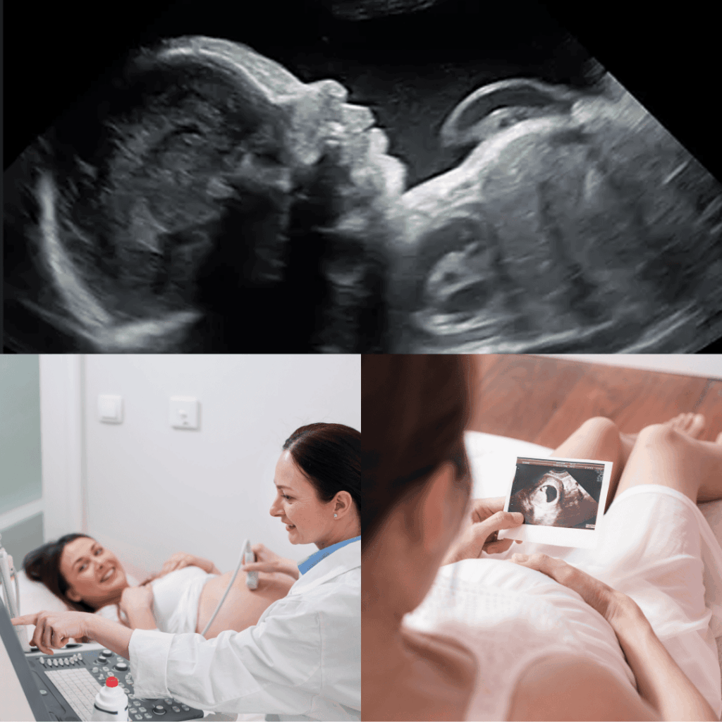
A maternity ultrasound scan uses gentle sound waves to create images of your baby during pregnancy. It offers parents the opportunity to see their baby, strengthen bonding, and capture meaningful moments throughout the pregnancy journey.
- Available in Klang Valley, Penang & Johor Bahru
- Digital scan images delivered to your Flabee Care App
What is the Meaning of a Maternity Scan?
A maternity ultrasound scan uses gentle sound waves to create images of your baby during pregnancy. It offers parents the opportunity to see their baby, strengthen bonding, and capture meaningful moments throughout the pregnancy journey.
Scans may be performed at different stages of pregnancy, including early scans to visualise the pregnancy and later scans to observe your baby’s growth and movements. An anatomy screening provides a general visual review of your baby’s physical development based on standard screening views, offering reassurance and a memorable experience for expectant parents.
All maternity ultrasound scans are conducted by trained professionals sonographer and are intended for screening and observation purposes only.
What Are the Best Weeks for an Ultrasound Scan?
The timing of an ultrasound scan is important to ensure clear visualisation and an optimal scanning experience at each stage of pregnancy.
- Dating Scan: The ideal time for a dating scan is between Weeks 8 and 10. At this stage, the pregnancy is more clearly visualised, allowing for a more reliable estimation of gestational age. Scans done earlier than 7 weeks may have limited visibility.
- Nuchal Translucency (NT) Scan: This scan is typically performed between Weeks 11 and 13+6. It is a time-sensitive screening that is only suitable within this specific window.
- Anatomy Scan Screening: An anatomy screening is usually scheduled between Weeks 18 and 22, with around Week 20 often considered ideal. At this stage, the baby’s structure and movements can be generally observed using standard screening views.
- Growth Scans: Growth scans may be performed in the third trimester, commonly from Week 28 onwards, to observe your baby’s size and position as the pregnancy progresses.
Pregnancy
Scan
From early pregnancy to 5D scans, experience maternity care enhanced by the Flabee Care App.


Frequently Asked Questions (FAQs) About Maternity Scans
1. Do I need a full bladder for the scan?
For early pregnancy scans (before 12 weeks), a full bladder is essential as it pushes the uterus up and provides a clearer “acoustic window.” For the 20-week scan, it’s often less critical, but always follow your clinic’s specific instructions.
2. Can I find out the baby's sex at the scan?
Some parents may start to have a general idea of the baby’s sex from around 15 weeks of pregnancy, depending on the baby’s position during the scan. At this stage, visibility may still be limited, and accuracy is not high (around 60%).
From around 20 weeks onwards, the baby’s anatomy is more developed and easier to visualise. If conditions are suitable, the accuracy of a general visual impression may increase to up to about 90%. However, this still cannot be guaranteed, as factors such as baby position and movement play an important role.
3. What if the scan detects a possible anomaly?
Try not to panic. Occasionally, an ultrasound image may show an appearance that requires further review or clarification. If this happens, you may be advised to follow up with your attending doctor, who may then refer you to a Maternal–Fetal Medicine (MFM) doctor if necessary.
An MFM doctor specialises in pregnancy care and may recommend additional scans or assessments to gain a clearer understanding. Your doctor will guide you through the next steps and provide appropriate advice and support.
Please note that our ultrasound scans are intended for screening and observation purposes only. They are not diagnostic, and any medical decisions should always be made in consultation with your doctor.
4. Why might I need a transvaginal scan?
In early pregnancy, a transvaginal (internal) scan may be recommended because it allows the probe to be closer to the uterus, which can provide clearer images compared to an abdominal scan at this stage. This can help with early visualisation when the pregnancy is still very small.
The scan is generally safe and is usually not painful, though some mild discomfort may be felt. It is performed by trained professionals with care and respect for your comfort and privacy.
Transvaginal scans are commonly used in early pregnancy and are intended for screening and observation purposes only.
5. My friend had more scans than me. Should I be worried?
No. The number of scans is tailored to each individual pregnancy. More scans are often required for monitoring specific conditions (e.g., twins, high blood pressure, gestational diabetes), not because it’s “better.” Fewer scans usually indicate a low-risk, progressing-as-expected pregnancy.
6. Can I have a copy of the ultrasound pictures?
Yes. At most ultrasound centres or sono clinic, you may request printed ultrasound images directly from the sonographer during your scan. Availability and charges may vary by clinic location, and an additional fee may apply.
Digital copies may also be available at selected locations. Where offered, images can be securely shared through the Flabee Care app for easy access and safekeeping.
Please feel free to let our sonographer know your preference during your appointment.
7. Can you help with insurance forms?
Insurance forms related to pregnancy or medical claims are not completed at our ultrasound centre. These forms are typically handled by your O&G specialist or attending gynaecologist, who oversees your medical care.
We recommend checking directly with your O&G doctor or clinic for assistance with insurance documentation.
8. Can I bring my family to the scan?
Yes, you’re welcome to bring your family along to share in this special moment. At most ultrasound centres, up to four accompanying persons are allowed during the scan, subject to space and clinic policy.
For everyone’s comfort and safety, we recommend checking with the ultrasound center or clinic in advance, as policies may vary by location.
9. What if my baby is in an unfavourable position during the scan?
This is very common, so there’s no need to worry. Babies often move or curl up in ways that make them shy on camera.
Our sonographer may gently guide you to change positions, ask you to take a short walk, empty your bladder, or lightly move your abdomen to encourage the baby to shift. These simple steps often help improve visibility during the scan.
If clear images are still difficult to obtain, the sonographer will advise you on the next best steps.
10. Why doesn’t my baby’s ultrasound photo look as clear as those in advertisements or on social media?
Ultrasound images can look very different from one pregnancy to another. The images you see in advertisements or on social media are usually selected from the clearest cases, taken under ideal conditions.
In real-life scans, image clarity can be affected by many normal factors such as the baby’s position, movements, stage of pregnancy, the amount of surrounding fluid, and individual body differences. Because every pregnancy is unique, it’s completely normal for images to vary in appearance.
Our goal is to provide you with the best possible images during your session, while ensuring a comfortable and meaningful experience. We encourage parents to enjoy the moment of seeing their baby rather than comparing images.
11. If I’ve done a scan at your centre, do I still need to follow up with my doctor?
Yes. Ultrasound scans performed at our centre are intended for screening and observation purposes only. They do not replace your regular pregnancy check-ups with your O&G doctor or attending healthcare provider.
Your doctor is responsible for your overall pregnancy care, including medical assessments, advice, and any necessary follow-up. We encourage all mothers to continue their scheduled appointments with their doctor, even after completing a scan with us.
12. Is ultrasound safe if I have it frequently?
Ultrasound has been used in pregnancy for many years and does not involve radiation. When performed by trained professionals and according to recommended practices, it is generally considered safe.
For many parents, having an ultrasound about once a month is commonly sufficient to visually observe the baby’s growth as the pregnancy progresses. However, in certain situations—such as early pregnancy, closer to the due date, or twin or multiple pregnancies—more frequent scans may be advised by your doctor to support closer monitoring.
We always recommend following the guidance of your O&G doctor, as they are best placed to advise on scan frequency based on your individual pregnancy.
13. What is the difference between an anatomy screening and a Detail scan?
An anatomy screening is a general ultrasound screening that looks at the baby’s overall physical structure using standard views. It is commonly offered as part of routine pregnancy scans to visually observe the baby’s development and movements. Anatomy screening is intended for screening and observation purposes only and does not involve in-depth analysis or diagnosis.
A Detail scan (sometimes referred to as a targeted or specialised scan) is usually performed in a hospital or specialist setting, often by a Maternal–Fetal Medicine (MFM) doctor or specialist. It involves a more comprehensive and focused assessment and is typically done when there is a medical indication or referral from an O&G doctor.
In short, an anatomy screening provides a general overview, while a Detail scan is a specialised examination carried out under medical supervision when needed.
14. Is an appointment required, or can I walk in?
An appointment is strongly recommended to help manage waiting time and ensure a smoother experience. Walk-in visits are not recommended, as availability depends on the clinic’s schedule and may involve longer waiting times.
Even with a scheduled appointment, waiting time may range from 30 to 60 minutes during peak hours. For parents who prefer minimal waiting time, a VIP package is available at selected locations, subject to availability.
15. What is the difference between 2D Full Anatomy Screening and 2D Partial Anatomy Screening?
2D Full Anatomy Screening
This screening is typically performed between 18 and 24 weeks of pregnancy, with around 20 weeks considered the optimal time. At this stage, the baby’s structure can generally be well visualised using standard screening views, allowing for a more comprehensive overview.
When conditions are suitable, overall screening visibility and accuracy may be up to about 90%, depending on factors such as baby position and individual differences.
2D Partial Anatomy Screening
This screening is commonly offered between 24 and 29 weeks of pregnancy, particularly for parents who may have missed the earlier recommended window. As the baby grows larger and has less space to move, full visualisation of all areas may be more limited.
As a result, overall screening visibility and accuracy are generally around 60–70%, depending on scan conditions.
Important note:
All anatomy screenings are provided for screening, observation, and bonding purposes only. They are not diagnostic scans and do not replace medical assessments, detailed scans, or follow-up care with an O&G doctor or Maternal–Fetal Medicine (MFM) specialist.