Everything You Need to Know Before Your X-Ray

An X-ray is a fast, painless imaging test that uses a small amount of high-energy electromagnetic radiation to produce images of the inside of the body. It is primarily used to visualize bones (which appear white) and diagnose conditions like fractures, infections, or tumors. Since its discovery by German physicist Wilhelm Conrad Röntgen in 1895, this form of electromagnetic radiation has transformed how physicians diagnose, monitor, and treat many medical conditions (World Health Organization, 2023).
Today, X-ray imaging is one of the most widely used diagnostic tools in modern medicine. Every year, millions of patients worldwide undergo X-ray examinations for a wide range of purposes—from routine dental assessments to emergency trauma evaluations (World Health Organization, 2023). According to the Ministry of Health Malaysia, radiological imaging plays a vital role in modern healthcare by enabling early detection, accurate diagnosis, and effective monitoring of medical conditions (Ministry of Health Malaysia, 2020).
This comprehensive guide aims to answer all these questions and more. Whether you are scheduled for an X-ray and want to know what to expect, or you are simply curious about this fascinating medical technology, we invite you to explore the complete picture of X-ray imaging—from its fundamental principles to what happens after the procedure is complete.
What is an X-Ray?
At its most fundamental level, an X-ray is a form of high-energy electromagnetic radiation that can penetrate body tissues and produce images of internal structures. X-rays exist on the electromagnetic spectrum between ultraviolet light and gamma rays, with wavelengths ranging from approximately 0.01 to 10 nanometers (World Health Organization, 2023).

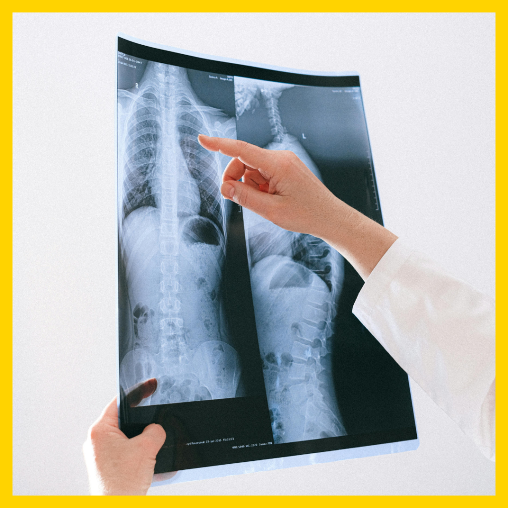
What makes X-rays particularly valuable in medicine is their ability to penetrate different tissues at different levels. Dense materials such as bones absorb more radiation, while softer tissues allow more X-rays to pass through. This difference in absorption creates contrast in the resulting image, allowing healthcare professionals to distinguish between bones, soft tissues, and air-filled spaces such as the lungs (Radiological Society of North America, 2023).
The resulting image shows bones as white or light grey, soft tissues as shades of grey, and air-filled spaces as darker areas. Radiologists—medical doctors specially trained to interpret medical images—analyze these patterns to identify injuries, abnormalities, or disease (World Health Organization, 2023).
Modern X-ray machines use advanced technology to generate and capture these images efficiently. According to the Ministry of Health Malaysia, medical imaging equipment must comply with strict safety and operational standards to ensure both accurate diagnosis and patient safety (Ministry of Health Malaysia, 2020).
Is There a Purpose for Doing an X-Ray?
The purposes of X-ray examinations are diverse and span nearly every medical specialty. At its core, an X-ray allows healthcare professionals to see inside the body without surgery, helping them answer clinical questions that cannot be resolved through physical examination alone (World Health Organization, 2023).
Diagnostic Purpose
The most common reason for performing an X-ray is to establish or confirm a diagnosis. For example, if a patient experiences a fall and cannot move a limb normally, an X-ray can reveal whether a fracture has occurred and determine its location and severity. This information is essential for guiding appropriate treatment (Ministry of Health Malaysia, 2020).
Monitoring Disease Progression
X-rays also play an important role in monitoring disease progression. For instance, chest X-rays may be used to monitor lung infections such as pneumonia or to assess fluid accumulation in patients with heart conditions. Repeated imaging allows doctors to track improvements or detect complications early (World Health Organization, 2023).
Guiding Treatment Decisions
In many situations, physicians require imaging before performing procedures or treatments. Dentists use X-rays before root canal procedures to assess the structure of teeth and surrounding bone. Orthopedic surgeons rely on X-rays to evaluate bone alignment before performing fracture repair or joint replacement surgery (Radiological Society of North America, 2023).
Screening for Early Detection
Certain X-ray examinations are used for preventive screening. For example, mammography is a specialized X-ray technique used to detect breast cancer at an early stage, sometimes before symptoms appear. Early detection significantly improves treatment outcomes and survival rates (World Health Organization, 2023).
Who Needs an X-Ray?
Determining who requires an X-ray involves careful clinical judgment. Although X-ray imaging provides valuable diagnostic information, it also exposes patients to ionizing radiation. Therefore, healthcare providers follow the ALARA principle (As Low As Reasonably Achievable) to ensure radiation exposure is minimized while still obtaining necessary diagnostic information (Ministry of Health Malaysia, 2020).
Trauma and Injury Patients
Individuals who experience significant trauma—such as motor vehicle accidents, sports injuries, or falls—may require X-rays to evaluate fractures, joint dislocations, or internal injuries (World Health Organization, 2023).
Individuals with Unexplained Symptoms
Patients experiencing persistent symptoms such as chest pain, chronic cough, unexplained abdominal discomfort, or joint pain may undergo X-ray examinations to identify possible underlying structural abnormalities (Radiological Society of North America, 2023).
Preoperative Patients
Before certain surgeries, physicians may request chest X-rays to evaluate lung health and heart size. This information helps assess the patient’s ability to tolerate anesthesia and identify any conditions that may complicate the procedure (Ministry of Health Malaysia, 2020).
Occupational Health Monitoring
Workers exposed to dust or hazardous environments—such as miners or construction workers—may undergo periodic chest X-rays to monitor lung health and detect occupational lung diseases (World Health Organization, 2023).
Special Populations
Certain groups may require specialized imaging. For example, infants may undergo X-ray examinations to assess developmental conditions, while elderly individuals may require imaging to detect fractures related to osteoporosis (Radiological Society of North America, 2023).
Pregnant women are generally advised to avoid unnecessary X-ray examinations. When imaging is unavoidable, protective shielding is used to reduce radiation exposure to the fetus (Ministry of Health Malaysia, 2020).
What Is the Inspection Procedure?
Understanding what happens during an X-ray examination can reduce anxiety and help patients cooperate effectively during the procedure.
Patient Registration and Medical History
The process typically begins with patient registration and verification of the examination request. Healthcare staff may ask about symptoms, medical history, and the possibility of pregnancy. This information helps ensure the correct imaging procedure is performed safely (Ministry of Health Malaysia, 2020).
Changing and Preparation
Patients may be asked to change into a hospital gown and remove any metal objects such as jewelry, glasses, or accessories. Metal can interfere with image quality and obscure important details (Radiological Society of North America, 2023).
Positioning
A radiographer or radiologic technologist positions the patient carefully to obtain clear images of the required body part. Proper positioning is essential for diagnostic accuracy and may require the patient to remain still for a short period (World Health Organization, 2023).
Image Acquisition
Once properly positioned, the technologist activates the X-ray machine from behind a protective shield. The exposure itself lasts only a fraction of a second, and most examinations require several images from different angles to provide a complete assessment (Ministry of Health Malaysia, 2020).
Image Review
After the images are taken, the technologist reviews them to ensure they are clear and diagnostically useful. If necessary, additional images may be taken before the patient leaves the department.
Why Is It Important to Have This X-Ray Examination?
The importance of X-ray examination extends far beyond simple curiosity about what lies beneath the skin. X-rays often provide information that fundamentally changes patient management and outcomes.
Establishing Accurate Diagnoses
Without X-ray imaging, many conditions would remain undiagnosed until they progressed to advanced stages. A hip fracture in an elderly patient might be mistaken for severe arthritis. A small lung cancer might grow silently until it causes symptoms. A dental abscess deep within the jawbone could spread infection throughout the body. X-rays reveal these hidden problems when they are most treatable.
Avoiding Unnecessary Treatment
Clinical examination alone can lead to incorrect assumptions. A child with a limp might have a hairline fracture requiring immobilization, but without an X-ray, they might be sent home with a diagnosis of a simple bruise. Conversely, what appears to be a fracture on physical examination might actually be severe soft tissue injury that requires completely different management. X-rays provide objective evidence that prevents both undertreatment and overtreatment.

Guiding Emergency Care
In emergency situations, X-rays provide rapid answers that guide life-saving interventions. A tension pneumothorax—air trapped in the chest cavity that compresses the heart and lungs—can be diagnosed within minutes of chest X-ray, allowing immediate placement of a chest tube. Bowel obstructions identified on abdominal X-rays prompt surgical consultation before perforation occurs.
Documenting Medical Conditions
X-rays create permanent records that serve multiple purposes beyond immediate clinical care. These images document the extent of injuries for workers’ compensation claims or personal injury lawsuits. They establish baselines against which future changes can be measured. They provide teaching material for medical education and research documentation for scientific studies.
Monitoring Treatment Effectiveness
Following treatment initiation, X-rays provide objective evidence of whether interventions are working. A pneumonia patient whose fever persists might need a follow-up X-ray to ensure the infection is responding to antibiotics. A cancer patient receiving radiation therapy undergoes periodic imaging to verify tumor shrinkage. An athlete recovering from a stress fracture returns for X-rays before being cleared to resume training.
What Preparations Are Needed to Perform This X-Ray Examination?
Preparation for X-ray examinations ranges from virtually none for simple extremity studies to more involved protocols for specialized examinations. Understanding these requirements helps ensure your examination proceeds smoothly.
General Preparations
For most routine X-rays—those of the arms, legs, chest, or spine—no special preparation is required. You may eat normally, take your regular medications, and continue your daily activities. You should wear comfortable clothing without metal fasteners when possible, though you will likely change into a gown regardless.
Pregnancy Considerations
If there is any possibility you might be pregnant, you must inform your physician and the radiology staff before the examination. This information is critical because developing fetuses are particularly sensitive to radiation exposure. In many cases, examinations can be postponed until after delivery or alternative imaging methods such as ultrasound may be substituted. When an X-ray is medically necessary during pregnancy, shielding will be used to minimize fetal exposure.
Barium Studies and Contrast Examinations
Some X-ray examinations require contrast materials to visualize specific structures. If you are scheduled for a barium swallow, barium enema, or intravenous pyelogram (kidney X-ray), specific preparation is necessary. Barium studies of the digestive tract may require fasting for 8-12 hours beforehand. Bowel preparation similar to that used for colonoscopy may be needed for lower gastrointestinal studies. Intravenous contrast studies require assessment of kidney function through blood tests beforehand.
Medication Management
Most routine medications can be taken as usual before X-ray examinations. However, if you are diabetic and require fasting for a contrast study, your diabetes medications may need adjustment. The radiology department will provide specific instructions if this applies to your situation.
What to Bring
When reporting for your X-ray examination, bring your physician’s order, insurance information, identification, and any prior X-ray images or reports if they were performed at another facility. Comparison with previous studies is often essential for accurate interpretation. Also bring a list of your current medications and any allergies, particularly allergies to contrast materials or iodine.
How Is an X-Ray Done?
Walking into an X-ray room for the first time can feel intimidating. The large machines, unfamiliar equipment, and clinical environment may seem overwhelming. Understanding exactly what happens during the procedure demystifies the experience and helps you cooperate fully.
Entering the Examination Room
The technologist will escort you to the examination room, which contains the X-ray machine—typically a large apparatus with a movable X-ray tube suspended from the ceiling or mounted on an articulated arm. Beneath or behind the X-ray tube is the image receptor, which captures the X-rays after they pass through your body. In digital systems, this receptor resembles a flat panel or plate.
Understanding the Equipment
The technologist may explain how the machine works and what you will experience. The X-ray tube will be positioned over or beside the body part being examined. A control panel behind a protective shield allows the technologist to adjust exposure settings and activate the machine while remaining safe from scattered radiation.
Assuming the Position
The technologist will guide you into the exact position needed for each image. For a chest X-ray, you will likely stand against a vertical receptor with your shoulders rolled forward and hands on your hips. For a knee X-ray, you may lie on the examination table with your leg positioned precisely. For a hand X-ray, you might place your palm flat on the image receptor while the X-ray tube is positioned above.
Breathing Instructions
For many examinations, particularly chest and abdominal X-rays, breathing instructions are crucial. You will be asked to take a deep breath and hold it. This accomplishes two things: it expands the lungs for better visualization, and it stops all motion. The technologist will watch you take that breath, then quickly retreat behind the shield and activate the machine before you need to exhale.
Multiple Images
Rarely is a single image sufficient for complete evaluation. Most body parts require at least two views taken at different angles. A standard chest X-ray includes front-to-back (posteroanterior) and side-to-side (lateral) views. An extremity examination might include front, side, and angled views. Each additional image requires repositioning, so the process repeats until all necessary views are obtained.
Special Considerations
Children require special attention during X-ray examinations. Pediatric technologists are skilled at explaining procedures in age-appropriate language and using distraction techniques. Parents may be allowed to stay with young children, typically wearing protective lead aprons. In some cases, gentle immobilization devices help children maintain position without repeated exposure.
Completion
Once all images are acquired and verified for quality, the examination is complete. The technologist will help you down from the table if needed and direct you to the changing area to dress. You are then free to leave unless you require additional imaging studies.
What Should You Expect After an X-Ray?
The immediate post-X-ray experience is typically uneventful, but understanding what happens next helps set appropriate expectations.
Immediate Sensations
X-rays themselves produce no sensation. You cannot feel them passing through your body, and there is no taste, smell, or other perception associated with the exposure. The procedure causes no pain—any discomfort comes only from holding positions that may be awkward or from lying on a firm table.
Return to Normal Activities
For routine X-rays without contrast material, you may resume all normal activities immediately. There are no restrictions on driving, eating, working, or exercise. The examination leaves no trace in your body, and you will feel exactly as you did before the procedure.
Contrast Material Considerations
If you received contrast material, specific post-procedure instructions may apply. Barium, if swallowed or administered as an enema, will eventually pass through your digestive tract and may cause white or light-colored stools for a day or two. Drinking extra fluids helps eliminate contrast from your system. Intravenous contrast is filtered by your kidneys and eliminated in urine, which may appear temporarily different.
Image Processing and Interpretation
In modern digital radiology departments, images appear on computer monitors almost instantly. However, immediate availability does not mean immediate interpretation. The technologist verifies technical quality, but the diagnostic interpretation must be performed by a radiologist—a physician with specialized training in image interpretation. This process typically requires several hours to a full day, depending on department workflow and examination urgency.
Receiving Results
Your X-ray results will be communicated to the physician who ordered the examination. This may be your primary care doctor, a specialist, or the emergency department physician. They will discuss the findings with you during a follow-up appointment, by phone, or through your electronic health record portal. Emergency departments typically provide results before discharge, while outpatient results may take several days to reach you.
Follow-Up Recommendations
Depending on what the X-ray reveals, your physician may recommend additional steps. Normal findings might provide reassurance and end the diagnostic process. Abnormal findings may prompt further imaging with other modalities, referral to specialists, or immediate treatment. Serial X-rays may be scheduled to monitor healing or disease progression over time.
Are There Any Side Effects or Risks That May Occur to the Patient?
The question of risks associated with X-ray examinations deserves careful, honest consideration. While X-rays are remarkably safe when used appropriately, they are not entirely without risk.
Radiation Exposure Concerns
The primary risk associated with X-rays stems from their use of ionizing radiation. This type of radiation carries enough energy to remove electrons from atoms, potentially damaging DNA within cells. The body has remarkable DNA repair mechanisms that usually correct this damage, but occasionally, unrepaired damage can lead to cancer years or decades later.
Quantifying the Risk
The radiation dose from a typical diagnostic X-ray is quite low. To put it in perspective:
– A chest X-ray exposes you to approximately 0.1 millisievert (mSv) of radiation
– This is equivalent to the background radiation you naturally receive from the environment over about 10 days
– A dental X-ray delivers even less—about 0.005 mSv
– Even a CT scan, which uses X-rays to create cross-sectional images, delivers only 2-10 mSv depending on the body part
The additional cancer risk from these doses is extremely small. One chest X-ray increases your lifetime cancer risk by approximately 0.0001%—a risk so tiny it is difficult to measure in population studies.
Pregnancy Risks
The developing fetus is more sensitive to radiation effects than adults. This sensitivity is highest during organ formation in the first trimester. However, even during pregnancy, necessary X-rays can be performed with appropriate precautions. Lead shielding placed over the abdomen and pelvis dramatically reduces fetal exposure. If an X-ray is truly needed for maternal health, the benefits almost always outweigh the minimal fetal risk.
Allergic Reactions
When contrast materials are used, allergic reactions become possible. Modern non-ionic contrast agents have greatly reduced reaction rates, but reactions still occur in approximately 0.2-0.7% of patients. Most reactions are mild—hives, itching, or nausea—but severe reactions including anaphylaxis can occur. Patients with known contrast allergies may receive premedication with steroids and antihistamines before future contrast studies.
Contrast and Kidney Function
Intravenous contrast is eliminated by the kidneys. Patients with severely reduced kidney function may be unable to clear contrast efficiently, potentially causing a condition called contrast-induced nephropathy. For this reason, kidney function is assessed through blood tests before contrast administration in at-risk patients. Alternative imaging methods may be substituted when kidney function is significantly impaired.
Repeat Examinations
The greatest risk from X-rays may come from unnecessary repeat examinations. When images are technically inadequate due to patient movement, improper positioning, or equipment malfunction, the examination must be repeated, doubling the radiation dose. Following technologist instructions carefully helps ensure first-time success.
Cumulative Exposure Concerns
While individual X-ray risks are minimal, cumulative exposure over a lifetime deserves consideration. Patients with chronic conditions requiring frequent imaging should discuss cumulative exposure with their physicians. Modern practice emphasizes using the lowest possible radiation dose that still provides diagnostic information—a principle called ALARA (As Low As Reasonably Achievable).
Balancing Risks and Benefits
The most important perspective on X-ray risks involves weighing them against the risks of not obtaining needed information. A missed fracture can lead to improper healing, chronic pain, and permanent disability. An undiagnosed pneumonia can progress to sepsis and death. Undetected cancer can spread beyond cure. In virtually all cases where X-rays are medically indicated, the benefits of accurate diagnosis dramatically outweigh the minimal radiation risks.
Conclusion
Since Wilhelm Röntgen first observed the mysterious rays that could see through flesh, X-ray imaging has evolved into one of medicine’s most versatile and valuable tools. From revealing hidden fractures to guiding complex procedures, from screening for breast cancer to monitoring pneumonia treatment, X-rays serve patients across virtually every medical specialty.
The technology has advanced remarkably—from glass photographic plates to instant digital images that can be shared around the world in seconds. Yet the fundamental principle remains unchanged: invisible rays create visible truth, allowing physicians to see what lies beneath the surface and make informed decisions about diagnosis and treatment.
For patients facing an X-ray examination, understanding the process transforms an unfamiliar experience into a predictable one. Knowing what to expect, why it matters, and how to prepare empowers you to participate actively in your healthcare. The minimal radiation exposure, the brief procedure time, and the immediate return to normal activities make X-ray one of the most patient-friendly diagnostic tools available.
The next time your physician recommends an X-ray, you can approach it with confidence rather than uncertainty. You understand now that this quick, painless procedure provides information that might otherwise remain hidden—information that could make the difference between appropriate treatment and missed diagnosis, between recovery and complication, between uncertainty and clarity.
In the end, that is the true purpose of every X-ray: to replace guesswork with knowledge, and to illuminate the path toward better health.

Get Clarity on Your Health—Book Your X-Ray Today
Don’t ignore symptoms or delay important diagnoses. A quick and painless X-ray can provide the answers you need to take the right next step for your health.
At Sonobee Ultrasound, we offer safe, reliable, and high-quality X-ray services performed by trained professionals using modern equipment—ensuring accurate results with minimal waiting time.
👉 Book your X-ray appointment with Sonobee Ultrasound today and get fast, trusted insights for better treatment and peace of mind.
10 Frequently Asked Questions About X-Rays
1. Does an X-ray hurt?
No, X-rays themselves are completely painless. You cannot feel them passing through your body. Any discomfort comes only from holding specific positions that may be awkward or from lying on a firm table. For patients with existing pain, the technologist will work with you to find positions that are as comfortable as possible while still obtaining quality images.
2. How long does an X-ray take?
The actual X-ray exposure lasts only a fraction of a second—less time than it takes to blink. However, the complete examination, including changing clothes, being positioned, and obtaining multiple views, typically takes 10-20 minutes. More complex studies may require additional time.
3. Can I eat or drink before an X-ray?
For routine X-rays of the arms, legs, chest, or spine, you may eat and drink normally. However, if your examination requires contrast material or involves the digestive tract, specific fasting instructions will be provided. Always follow the preparation instructions given by your physician or the radiology department.
4. What should I wear for an X-ray?
Wear comfortable clothing without metal fasteners when possible. Buttons, zippers, snaps, and underwire bras contain metal that can appear on X-ray images. You will likely be asked to change into a hospital gown regardless of what you wear, so dressing in easily removable clothing is practical.
5. Can I have an X-ray if I'm pregnant?
If you are pregnant or suspect you might be, you must inform your physician and the radiology staff before any X-ray. In many cases, examinations can be postponed or alternative imaging methods used. When an X-ray is medically necessary during pregnancy, shielding will be used to protect the developing baby.
6. Will I be exposed to radiation from an X-ray?
Yes, X-rays do involve exposure to ionizing radiation. However, the amount is quite small—comparable to the natural background radiation you receive from the environment over days to weeks. The benefits of accurate diagnosis typically far outweigh these minimal risks.
7. How soon will I get my X-ray results?
The technologist who performs your X-ray cannot provide results—only a radiologist or your physician can interpret the images. Results are typically available to your doctor within 24-48 hours. In emergency settings, preliminary results may be available much sooner.
8. Can someone stay with me during the X-ray?
In most cases, yes. Family members or companions may accompany you, particularly if you need assistance or are anxious. They will be required to wear a lead protective apron if they remain in the room during exposure. Pregnant companions should wait outside the examination room.
9. Why do I need to hold my breath?
Breathing causes movement, which blurs X-ray images just as it blurs photographs. Holding your breath stops this motion, ensuring sharp, clear images. For chest X-rays, holding your breath after a deep breath also expands your lungs, providing better visualization of lung tissue.
10. Are digital X-rays different from traditional X-rays?
Digital X-rays use electronic sensors instead of photographic film to capture images. They require less radiation than traditional film X-rays, produce images that can be enhanced and shared electronically, and eliminate chemical processing. However, the basic principle and patient experience remain very similar.
Disclaimer: The information provided in this article is for educational purposes only and does not constitute medical advice; please consult a qualified healthcare professional for any medical concerns or before undergoing any radiological procedure.